Back to Profile Page

Tree of Life Media Contributed By Brian S. Leander

<< < 1 2 3 4 5 6 7 > >>
ID Thumbnail Media Data
32114
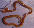
Scientific Name Thelepus sp.
Comments host of intestinal gregarines
Specimen Condition Live Specimen
Image Use creative commons This media file is licensed under the Creative Commons Attribution-NonCommercial License - Version 3.0.
Copyright © Brian S. Leander
Attached to Group Terebellida (Annelida): view page image collection
Gregarina: view page image collection
Title thelepussp.jpg
Image Type Photograph
Image Content Specimen(s)
ID 32114
32117
Scientific Name Lumbrineris sp.
Comments host of intestinal gregarines
Specimen Condition Live Specimen
Image Use creative commons This media file is licensed under the Creative Commons Attribution-NonCommercial License - Version 3.0.
Copyright © Brian S. Leander
Attached to Group Eunicida (Annelida): view page image collection
Gregarina: view page image collection
Title lumbrinerissp.jpg
Image Type Photograph
Image Content Specimen(s)
ID 32117
32126
Image Use creative commons This media file is licensed under the Creative Commons Attribution-NonCommercial License - Version 3.0.
Copyright © 2008 Brian S. Leander
Attached to Group Euglenida: view page image collection
Title euglenidstree.png
Image Type Diagram
ID 32126
32131
Comments A phagotrophic cell with >40 thin pellicle strips that are helically arranged and capable of metaboly.
Copyright © Brian S. Leander
Image Use ToL use only
Attached to Group Euglenida: view page image collection
Title phagotrophiceuglenid40.jpg
Image Type Photograph
Image Content Specimen(s), Ultrastructure
Technical Information Light micrograph
ID 32131
32132
Comments Photoautotrophic cell showing chloroplasts and the stigma.
Copyright © Heather Esson and Brian S. Leander
Image Use ToL use only
Attached to Group Phacus: view page image collection
Title photoautotrophiceuglenid2.jpg
Image Type Photograph
Image Content Specimen(s), Ultrastructure
Technical Information Light micrograph
ID 32132
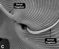
32135
Specimen Condition Dead Specimen
Body Part two flagella emerging from the anterior opening of the flagellar pocket
Image Use creative commons This media file is licensed under the Creative Commons Attribution-NonCommercial License - Version 3.0.
Copyright © 2008 Brian S. Leander
Attached to Group Peranema trichophorum (Peranema): view page image collection
Title peranema_trichophorum_flage.jpg
Image Type Photograph
Image Content Ultrastructure
Technical Information Scanning electron micrograph
ID 32135
32136
Comments Organization of the microtubular roots and paraxial rods associated with the euglenid flagellar apparatus.
Reference modified from Leander, B. S. 2004. Did trypanosomatid parasites have photosynthetic ancestors? Trends Microbiol. 12:251-258.
Image Use creative commons This media file is licensed under the Creative Commons Attribution-NonCommercial License - Version 3.0.
Copyright © 2008 Brian S. Leander
Attached to Group Euglenida: view page image collection
Title flagellarapparatus.jpg
Image Type Diagram
Image Content Ultrastructure
ID 32136
32138
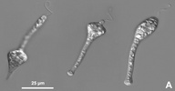
Location Specimen collected from a freshwater aquaculture pond near Boise, Idaho
Comments Serial images of the colorless euglenid flagellate, Astasia klebsii (Lemmermann, 1910) showing its characteristic euglenoid movement (metaboly).
Specimen Condition Live Specimen
Source Astasia klebsii
Source Collection Micro*scope
Image Use creative commons This media file is licensed under the Creative Commons Attribution-NonCommercial-ShareAlike License - Version 2.5.
Copyright ©
Creation Date January 2004
Attached to Group Astasia klebsii (Astasia): view page image collection
Title astasiamovement.jpg
Image Type Photograph
Image Content Specimen(s)
Technical Information DIC optics
ID 32138
32137
Comments Organization and main components of the euglenid pellicle: illustration shows the isolated proteinaceous strip and the complexity of the prearticular projections in many photoautotrophic euglenids.
Reference Modified from Leander, B. S., H. J. Esson, and S. A. Breglia. 2007. Macroevolution of complex cytoskeletal systems in euglenids. BioEssays 29:987-1000.
Creator Heather Esson and Brian Leander
Copyright © 2007 Heather Esson and Brian Leander
Image Use restricted
Attached to Group Euglenida: view page image collection
Title strip2.jpg
Image Type Diagram
Image Content Ultrastructure
Subject Histology, Morphology
ID 32137
32140
Comments Organization of the euglenid feeding apparatus: transverse view of the microtubule-reinforced feeding rods and vanes.
Copyright © Brian S. Leander
Image Use ToL use only
Attached to Group Euglenida: view page image collection
Title feedingapparatustem1.jpg
Image Type Photograph
Image Content Ultrastructure
Technical Information transmission electron micrograph
ID 32140
Please note: Most images and other media displayed on the Tree of Life web site are protected by copyright, and the ToL cannot act as an agent for their distribution. If you would like to use any of these materials for your own projects, you need to ask the copyright owner(s) for permission. For additional information, please refer to the ToL Copyright Policies.

Contribute to the ToL

ToL Contributions

Ways to Contribute

Use of Contributions

Scientific Content

Building Treehouses

Images/Media

  go to the Tree of Life home page
top